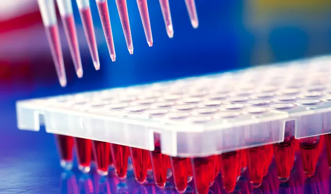
qPCR Probes

Home » Product & Services » Oligo Synthesis » qPCR Probes
Overview
qPCR Probes are important raw materials in qPCR. This technology has been widely used in scientific research and production practice, especially in medical testing and diagnosis.

Tsingke has a cutting-edge 100,000-level clean room, adhering to stringent biomedical diagnostic standards. Leveraging state-of-the-art synthesis technology, premium-quality synthesis reagents, advanced automated production equipment, and a rigorous ISO 13485 quality control system.

Our comprehensive range of qPCR probes includes various types such as qPCR Probes, MGB Probes, Double-Quenched Probes, Molecular Beacons and more. Tsingke could provides top-notch customized qPCR probes and oligo primers for qPCR experiments and kit. 
Advantages
Customer Trust
Serving over 500 IVD companies

Stable product quality
High-quality synthetic raw materials and technology, the probes have high purity and low background noise
Anti-contamination Procedures
Strictly control contamination from E.coli and human sources to avoid NTC peaks

Service Details

Service Name

Length (nt)

Purification

Price/
Turnaround time

Deliverable

Application

qPCR Probes 

15-30

DSL/OPC/PAGE/HPLC/
Dual PAGE & HPLC

Inquire

· Tube or Customized

· Lyophilized DNA

·COA Report
(electronic)

The most commonly used types of qPCR experiments

MGB Probes

13-25

For qPCR experiments with higher TM 

Double-Quenched Probes

15-45

For qPCR experiments with longer probes

Molecular Beacons

25-40

For qPCR experiments with extremely high sensitivity requirements

Other Probes

Customized

Customized

Special application directions

*Note: In addition to the recommended content, Oligo length and purification methods can also be customized.                                     
                 
Common Fluorescent Dyes and Quenchers types 

Fluorescent Dyes

Max Abs

Max Em

Quenchers

Quenching range

Quenching Max

FAM

494 nm

518 nm

Dabcyl

380 nm-530 nm

452 nm

TET

521 nm

536 nm

Eclipse

390 nm-625 nm

522 nm

JOE

520 nm

548 nm

MGB

390 nm-625 nm

522 nm

VIC

538 nm

554 nm

TAMRA

470 nm-560 nm

544 nm

HEX

535 nm

556 nm

BHQ1

480 nm-580 nm

534 nm

Quasar 570

547 nm

570 nm

BHQ2

550 nm-650 nm

579 nm

Cy3

552 nm

570 nm

BHQ3

620 nm-730 nm

672 nm

TAMRA

565 nm

580 nm

ROX

585 nm

605 nm

Texas Red

595 nm

615 nm

Alexa Fluor 633

632 nm

647 nm

Cy5

643 nm

667 nm

Quasar 670

647 nm

667 nm

Cy5.5

684 nm

710 nm

Cy7

750 nm

773 nm


Workflow
high-throughput synthesizer from pmol to mmol levels
Synthesis
high-throughput synthesizer from pmol to mmol levels
waters 2695 & Waters 2767 automatic purification and PAGE purification
Purification
waters 2695 & Waters 2767 automatic purification and PAGE purification
all MASS quality inspection, CE and other additional quality inspections
QC
all MASS quality inspection, CE and other additional quality inspections
automatic dispensing instrument for accurate dispensing
Distribution
automatic dispensing instrument for accurate dispensing
tube or Customized、Lyophilized DNA、COA Report
Delivery
tube or Customized、Lyophilized DNA、COA Report
Case 
Figure 1: qPCR Probe for SNP target gene quantification
Figure 2: MGB Probe for SNP detection


Figure 3: Long-term stability


Figure 4: Short-term stability


Figure 5: Fluorescence modification stability (70 H)
Related Resource
FAQ
What are the most commonly used fluorescent dyes?
FAM
TET
HEX
TAMRA
ROX
Cy3
Cy5
What causes low fluorescent signal in amplification curves?
First check the baseline-corrected or ROX-corrected raw curves. Low signal values in the amplification curves are mostly due to high background fluorescence. In probe-based detection, high background fluorescence is often caused by poor probe design, leading to insufficient quenching by the quencher group, improper pairing of the reporter and quencher groups, or low fluorescence labeling efficiency of the probe.
What causes erratic peaks in melting curves?
Erratic peaks in melting curves can result from contamination in the reaction system. Confirm contamination using NTC and NRC results, and check potential sources such as water, oligos, enzymes, and the experiment settings. Reagents exposed to strong light or high temperatures may degrade, so it is recommended to compare results with fresh reagents.
How to test the purity of primers?
A common method used in laboratories is the PAGE. For electrophoresis, use a polyacrylamide gel with 7 M urea, 20% polyacrylamide gel for primers with less than 12 bases, 16% polyacrylamide gel for primers with 12-60 bases, and 12% polyacrylamide gel for primers with more than 60 bases. Take 0.2-0.5 OD primer, dissolve with urea saturated solution or add urea dry powder into primer solution until saturated, and heat denaturation (95 ℃, 2 min) before loading sample. The purpose of adding urea is to denaturate, and to increase the specific gravity of the sample, which is easy to add the sample. After a certain period of time (about 2-3 hours), strip the polyacrylamide gel and detect the band type with a fluorescent TLC plate under the UV lamp. There is no impurity under the main band, indicating that the purity is good.
(Sometimes due to insufficient denaturation, there may be bands above the main band, which are primer secondary structure bands.)
Why do dissolved primers work fine at first but not after a period of time?
 If the water in which you dissolve the primers has a low pH or is contaminated with bacteria or nucleases, the primers will degrade. Inadequate thawing and mixing during use, and uneven liquid may also cause inaccurate amounts of primers. It is recommended to aliquot primers, avoid repeated freezing and thawing, and use 10 mM Tris pH 7.5 buffer to dissolve primers. There is another possibility that there is no problem with the primers, but that the quality of the materials used in PCR, especially the template, is not completely consistent with that used previously.
Will the primers degrade when transported at room temperature?
It will not degrade, and the dry primer can be stably stored at room temperature for at least two weeks. The average transport time is usually 5days, so the primers you receive will not degrade.
How to measure the OD value of primers?
The OD value of the synthetic primer was determined by using an ultraviolet spectrophotometer with a wavelength of 260 nm and a quartz colorimetric cup with an optical path of 1 cm to determine the optical density of the solution. The optical density of the solution should be diluted to 0.2-0.8. After the DNA dry powder is fully oscillated and dissolved with a certain volume of water, the OD value is measured by diluting with 1 mL of water. OD value of mother liquor should be converted according to dilution ratio. For example, to verify whether the amount of 2 OD primer is accurate, the simple practice is to add 1 mL water, thoroughly dissolve and mix, take 100 μL, add 900 μL water, a quartz colorimetric cup with a light diameter of 1 cm, a wavelength of 260 nm, and the absorbed reading at this time is 0.2.
How many primers of OD value should be synthesized?
According to the purpose of the experiment. In general, about 20 bases of primer 2 OD in PCR amplification can do 400 rxns standard PCR reactions of 50 μL system. If it is to do gene splicing or annealing to do the ligation, 1 OD is enough. 
**For Research Use Only. Not for use in diagnostic procedures.
Send Your Request
Want to learn more about these services? Speak to our experts and get a free quote.
Those with * are required